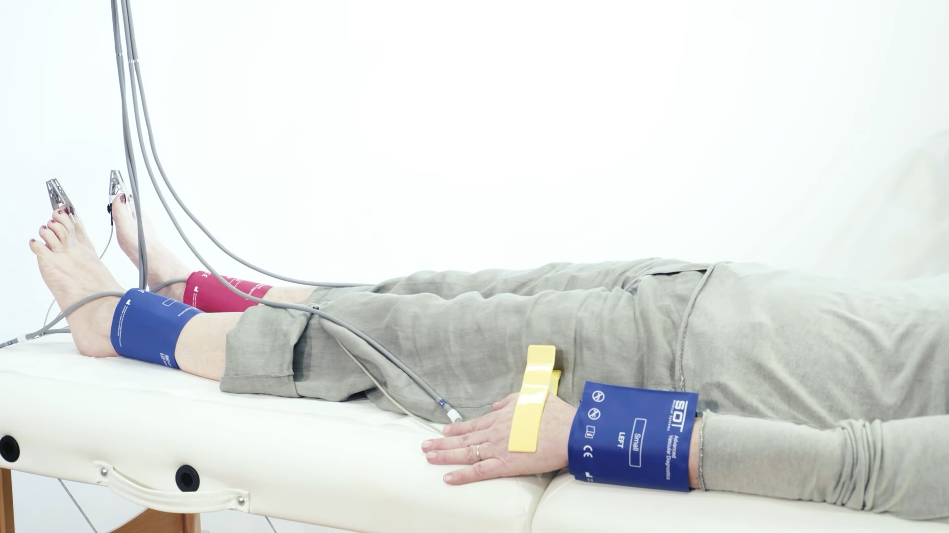
TOPP-Method (Tissue Optical Perfusion Pressure)
Description
TOPP-ABI includes simultaneous automated oscillometric ABI and PWI determination together with the systolic tissue optical perfusion pressure (TOPP) at the far end of microperfusion at the toe in a single-shot process. This standardized technique provides a new insight into foot artery and micro-hemodynamics on top of routine ABI measurements.
“Critical limb ischemia with consecutive foot ulcers and minor amputations are below the measuring thresholds of ABI. Since peripheral interventions enter the region of the pedal arteries, additional criteria determining the improvement of macrocirculatory and microcirculatory hemodynamics are of utmost interest.”
Tissue optical perfusion pressure: a simplified, more reliable, and faster assessment of pedal microcirculation in peripheral artery disease; Horstick, Messner, Grundmann, Yalcin, Weisser, Espinola-Klein; Vol. 319: H1208–H1220, 2020, AJP Heart – American Physiological Society
TOPP provides information of pedal macrocirculation and microcirculation, which cannot be achieved by ABI analysis alone.
With TOPP plus oscillometric ABI (TOPP-ABI) used as a one-stop-shop screening method, patients with unknown or unconfirmed PAD are sooner diagnosed and treated earlier to reduce disease progression.
Measurement Principle
After applying the cuffs on ankles and wrists, as well as the optical sensors on the toes, the system applies a pressure of 180mmHg to all cuffs, that is decreased by steps of 10mmHg.
By recording the optical sensors, the examiner can immediately determine the pressure step at which the patient’s toes show the first pulsations. Different key indicators, like the oscillometric ABI, the PWI, the amplitude or the rise time of the pulse wave, are recorded simultaneously.
In addition, important indicators for pedal blood circulation, such as the toe waveforms, are collected within the same measurement.

In addition, the TOPP measurement procedure often includes an exercise stress test to further clarify the results. This is because an asymptomatic arterial lesion in the lower limbs may become hemodynamically relevant after induced stress. The procedure is used to differentiate between vascular disorders and their severity.
Main Parameters
| Parameter | Name | Description |
|---|---|---|
| PWI | Pulse Wave Index | Maximal amplitude of the upper extremity (wrists) divided by the maximal pulse amplitude measured at the respective lower leg (ankles) and then multiplied by the respective ankle rise time of the pulse wave. Values above 220 are considered abnormal. |
| oABI | Oscillometric Ankle-Wrist-ABI | Mean arterial pressure of the ankle related to the mean arterial pressure of the wrist; values ≤ 0.9 indicate a vascular disorder (PAD). |
| HRV | Heart-Rate- Variability | Heart rate variability (HRV) is the physiological phenomenon of variation in the time interval between heartbeats. It is measured by the variation in the beat-to-beat interval. The ideal HRV varies over age and is individual to each patient. |
| PWV | Pulse-Wave-Velocity | PWV is the velocity at which the blood pressure pulse propagates through the circulatory system and should generally be ≤10 m/s. It is used clinically as a measure of arterial stiffness. |
| TIME SHIFT | ms | The time shift, measured between the start point of both pulse curves, shows the propagation time difference between the left and the right extremity. Values above 50 milliseconds are considered abnormal. |
| AMPLITUDE | mmHg | The height from the beginning of the steepest rise to the highest point of the pulse curve is the amplitude. |
| RISE TIME | ms | The rise time marks the time interval of the steepest rise to the highest point of the pulse curve. It should be < 200 milliseconds for normal values. |
| RISE TO FALL | % | The quotient, stated in percentage, of rise time and fall time. It should be < 33% for normal values. |
| OSCILLOMETRIC INDEX | mmHg | The oscillometric index marks the pressure stage where the highest amplitude was measured and is comparable to the mean arterial pressure. Values higher than 120 mmHg are considered abnormal. |
| SKIN TEMP | Skin Temperature | Skin temperature on toes recorded by the probes on the optical sensors. Side differences of more the 3 °C should be observed. |
Medical Applications
- Patients at increased risk of Lower Extremity Peripheral Artery Disease (PAD) and/or Diabetic Foot:
- Aged ≥ 65 years
- Diabetes, Smoking, Hyperlipidemia, Hypertension, and other risk factors for atherosclerosis
- Family history of PAD or other known forms of atherosclerosis
- Patients with medical conditions in the legs
- Monitoring of vascular status pre- and post-intervention
Test Examples
Table of Contents


