PVR – Pulse Volume Recording
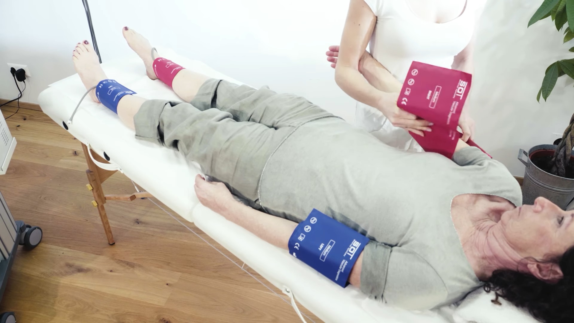
Pulse Volume Recording describes a further development of the traditional Oscillography measurement principle. By applying and inflating pneumatic measurement cuffs on specific positions of the extremities, like upper arms, wrists, thighs, calf, ankles, fingers or toes, the system can detect the pressure changes caused by the blood flow. Modern devices are sensitive enough to record the pulse wave form (including its relevant characteristics, such as amplitude, rise time, dicrotic notch, etc.) and not just the oscillations provoked by the patients’ cardiac cycle.
The standard test procedure required the examiner to apply pairs of cuffs (left and right) on 4, 6, or 8 measurement positions. The device will then simultaneously inflate all cuffs to a predefined starting pressure (e.g., 180 mmHg) and record the provoked pulse oscillations for a specific duration (e.g., 10 seconds). Consequently, the pressure on all cuffs will be reduced simultaneously to reach the next pressure step (e.g., 170 mmHg) and to record the pulse oscillations accordingly. This routine ends once the predefined minimum measurement value is reached, typically at 20 mmHg.
Although this test is mostly qualitative in nature, and the focus is on the shape of the obtained pulse wave forms, the system will determine different quantitative values, such as the Oscillometric Index, the maximal amplitudes, and the rise times for each measurement track. Based on the measurement setup, the system may also calculate ABI and PWITM.
Main Parameters
| Parameter | Unit / Description | Description |
|---|---|---|
| AMPLITUDE | mmHg | The height from the beginning of the steepest rise to the highest point of the pulse curve is the amplitude. |
| RISE TIME | ms | The rise time marks the time interval of the steepest rise to the highest point of the pulse curve and should be < 200 milliseconds. |
| RISE TO FALL | % | The quotient, stated in percentage, of rise time and fall time. It should be < 33% for normal values. |
| TIME SHIFT | ms | The time shift, measured between the start point of both pulse curves, shows the propagation time difference between the left and the right extremity. Abnormal: Values above 50 milliseconds. |
| PWV | Pulse-Wave- Velocity | PWV is the velocity at which the blood pressure pulse propagates through the circulatory system and should generally be ≤10 m/s. It is used clinically as a measure of arterial stiffness. |
| PWI | Pulse Wave Index | Maximal amplitude of the upper extremity (wrists) divided by the maximal pulse amplitude measured at the respective lower leg (ankles) and then multiplied by the respective ankle rise time of the pulse wave. Values above 220 are considered abnormal. |
| oABI | Oscillometric Ankle-Wrist-ABI | Mean arterial pressure of the ankle related to the mean arterial pressure of the wrist; values ≤ 0.9 indicate a vascular disorder (PAD). |
| OSCILLOMETRIC INDEX | mmHg | The oscillometric index marks the pressure stage where the highest amplitude was measured and is comparable to the mean arterial pressure. Values higher than 120 mmHg are considered abnormal. |
Medical Applications
- Differential clarification of Peripheral Artery Disease (PAD)
- Suspicion of Intermittent Claudication or Limb Ischemia
- Patients with medical conditions in the legs, such as:
- Swollen legs
- Skin alterations
- Cold or numb legs
- Short walking distance or pain at rest
- Skin lesions on ankle , feet or toes
- Patients at increased risk of lower extremity Peripheral Artery Disease (PAD):
- Aged ≥ 65 Years
- Diabetes, Smoking, Hyperlipidemia, Hypertension and other risk factors for atherosclerosis
- Family history of PAD or other know forms of atherosclerosis
- Monitoring of vascular status pre- and post-intervention
4 Channel Oscillography
Measurement Principle
The 4-channel oscillography test is typically performed on the lying patient on wrists and ankles. This is because their similar diameter allows for the comparison of pulse amplitudes.

The patient is instructed not to move or speak during the test to prevent measurement artefacts. After the procedure starts, the cuffs will inflate and reduce their pressure by the defined steps.
Depending on the settings, the measurement will be completed after two or three minutes. The resulting oscillograms should be assessed with a special focus on side differences of pulse wave forms and parameters.
Test Examples
Normal 4-Channel Oscillography Test
 |
Wrist and Ankle Pressure Tracks |
| Result Table with PWI, oABI, Amplitudes and Rise Times | |
| Mean Pulse Waves and Amplitude Histogram |
| Interpretation Criteria | |||||
| Side Difference | The mean arterial pressure of left and right side is almost the same, as well as the amplitude height. | ||||
| Pulse Wave | The rises of all pulse waves ascend steeply, and the heights of the amplitudes are good. The peaks of the pulse waves are normal. A dicrotic wave is visible and the pulse waves decrease not too steep and not too flat. This results in a good PWI™, which takes the rise time and the height of the amplitude into account. | ||||
| PWI™ | < 180 | Time Shift and Rise Time | Both within reference range. | Pulse Wave Velocity | ≤ 10 m/s |
| oABI | > 0.9 |
Pathologic 4-Channel Oscillography Test
 |
Wrist and Ankle Pressure Tracks |
| Result Table with PWI, oABI, Amplitudes and Rise Times | |
| Mean Pulse Waves and Amplitude Histogram |
| Interpretation Criteria | |||||
| Side Difference | There is a significant difference between the highest mean arterial pressures of the left and right side. The pressure of the left leg is much lower than the pressure of the right side, as well as the amplitude. Furthermore, the pressure of the left leg compared to the pressure of the left arm is much lower. Additionally, the highest amplitudes are at very high cuff pressures, which could indicate that the patient suffers from an arterial hypertension (at the arm 140mmHg) or incompressible arteries (media sclerosis). The oABI value on the left is pathologic. On the right side it shows that the blood flow in the right extremities is sufficient. | ||||
| Pulse Wave | Very small pulse waves in the left lower extremity, which could be an indication of a compensated occlusion. The dicrotic wave on the right side is visible in the lower position, but the height of the amplitude is slightly lower than in the arms which leads on to a bad PWI™. Considering that the patient had a vascular intervention on the right side, the PWI™ is acceptable in this case. The PWI™ on the left reacts to the inadequate values. | ||||
| PWI™ | > 220 | Time Shift and Rise Time | The time shift is in a regular range but the rise time on CH3 is too high. | Pulse Wave Velocity | Since the results show a blood flow disorder, PWV should not be considered, as it might be influenced. |
| oABI | L: <0.9(R :> 0.9) |
8 Channel Segmental Oscillography
Measurement Principle
An 8-channel segmental oscillography is usually performed simultaneously on wrists, thighs, calfs and forefoots on both sides. This allows for a comprehensive screening of the arterial blood flow in the lower extremities and presents a first indication for a possible occlusion and its position.

The patient is instructed not to move or speak during the measurement to prevent pulse wave artefacts. After the procedure starts, the cuffs will inflate and reduce their pressure by the defined steps.
Depending on the settings, the measurement will be completed after two or three minutes. The resulting oscillograms should be assessed with a special focus on side differences of pulse wave forms and parameters.
As with all PVR tests, the measurement can be performed at one or at multiple decreasing pressure steps. In case of the latter, the system will also calculate the oABI and Oscillometric Index in addition to several other pulse wave parameters.
Test Example
Table of Contents


