VOP – Venous Occlusion Plethysmography
Description
The venous vascular system is characterized by its large capacity. It contains about two thirds of the total amount of blood. In addition, veins are much more flexible than arteries. The veins have a valve mechanism, the so-called venous valves, so that the backflow of blood from the periphery can be guaranteed even in an upright posture.
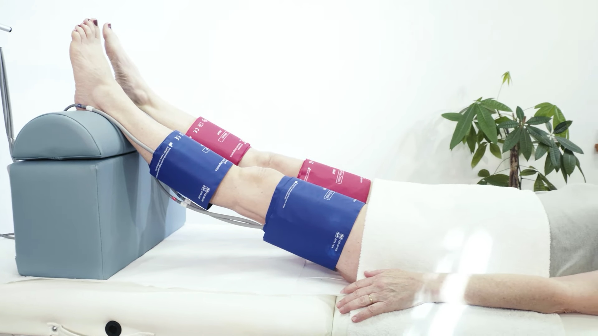
When the patient is lying down and the legs are raised well above the level of the heart, the leg veins run almost completely empty of pressure passively. Only the amount of blood that is maintained by the arterial inflow runs through the veins.
If the venous outflow of the leg is blocked on the thigh, the arterial inflow leads to an increase in the volume of blood that collects in the leg veins with each heartbeat. Due to the large volume of the veins, it takes several minutes for the leg veins to be filled completely.
The venous capacity is a rather theoretical value. It is the maximum volume of blood that the peripheral veins can hold. The current vein volume can be influenced by the ambient temperature and the state of hydration as well as by varicosis or thrombosis. Therefore, this test helps to determine or exclude these conditions.
Measurement Principle
While performing a VOP test, cuffs are applied on thighs and calves. The legs of the patient are elevated over heart height. The thigh cuffs inflate up to 80mmHg to prevent the venous outflow but not to affect the arterial inflow of blood. At the same time, the calf cuffs act as highly sensitive sensors.

The measurement procedure takes three minutes, during which the leg veins increase in volume. After this time, the system immediately releases the cuff pressure on the thighs. With a healthy patient, this leads to a rapid outflow of the retained venous blood.

Main Parameter

| Parameter | Unit | Description | Reference Value |
|---|---|---|---|
| Venous Capacity VC | ml/100 ml | Is defined as the venous increase from the start value to the end value after 3 minutes. | 3 – 6 ml/100 ml |
| Venous Outflow V0 | ml/100 ml/min | Is represented by the line drawn through point A and B. Point A is fixed at 500ms after opening of the compression cuffs. Point B is positioned 1000ms later than point A. This line is defined in ml/100 ml/min. | ≥ 30 ml/100 ml/min |
| Arterial flow | ml/100 ml/min | This indicator is also displayed on the results table and expresses the relative velocity of the volume increase. | Not defined |
Medical Applications
- Suspicion of Vein Thrombosis (pelvic, legs or arms)
- Suspicion of Post-Thrombotic Syndrome (PTS)
- Suspicion of Varicosis
- Clarification of patients with swollen legs
- Monitoring of vascular status pre- and post-intervention
Test Examples
Table of Contents

