Author: Miguel Marhl

Guide on Effective PAD Screening

Guide on Effective PAD Screening

Download the comprehensive guide with focused contents on PAD screening: Peripheral Artery Disease (PAD) Symptoms of PAD Treatment of PAD How to assess PAD The traditional method: Ankle Brachial Index (ABI) The modern approach: Pulse Wave Index (PWI™) Vascular Screening with the TOPP-Method Measurement Principle Obtained Parameters Interpretation Example Case Study with Angiography Bonus: Exercise […]

Read More
VOP – Venous Occlusion Plethysmography

VOP – Venous Occlusion Plethysmography

The venous vascular system is characterized by its large capacity. It contains about two thirds of the total amount of blood. In addition, veins are much more flexible than arteries. The veins have a valve mechanism, the so-called venous valves, so that the backflow of blood from the periphery can be guaranteed even in an […]

Read More
D-PPG Muscle Pump Test (Venous Reflux)

D-PPG Muscle Pump Test (Venous Reflux)

The Venous Reflux Test (with D-PPG – Digital Photoplethysmography) is a non-invasive screening method for the functional assessment of the hemodynamics of the leg vein system and to determine the competence of the venous valves. Insufficient competence of the superficial venous valves, for example, may require the patient to wear compression stockings for compensation. By attaching […]

Read More
TOS-Test (Thoracic Outlet Syndrome)

TOS-Test (Thoracic Outlet Syndrome)

Thoracic outlet syndrome (TOS) is a condition in which there is compression of the nerves, arteries, or veins in the passageway from the lower neck to the armpit. Usually, extrinsic factors are responsible for the symptoms of TOS, but an aneurysm of the subclavian artery can also trigger TOS. Intrinsic factors, such as activities that […]

Read More
Exercise Stress Test

Exercise Stress Test

Several guidelines recommend an exercise stress test to further clarify results. This is because an asymptomatic arterial lesion in the lower limbs may become symptomatic after induced stress. Essentially, the procedure is used to differentiate between vascular disorders and their severity. Exercise stress tests can be performed in different ways together with TOPP, PVR or […]

Read More
OAP – Optical Arterial Pressure

OAP – Optical Arterial Pressure

Optical Arterial Pressure is a measurement method to determine systolic blood pressure in either finger or toes. The Trans-Atlantic Inter-Society Consensus Document on Management of Peripheral Arterial Disease (TASC) recommends the determination of the systolic toe pressure for diagnosis of CLI (critical limb ischemia). It is also valuable in assessment of severe peripheral artery disease, […]

Read More
OPO – Optical Pulse Oscillography

OPO – Optical Pulse Oscillography

Similar to the pneumatic Pulse Volume Recording (PVR), oscillations of the pulse waves can be recorded using optical photoplethysmography (PPG) sensors. By emitting infrared light, these probes can detect variations in the microcirculatory blood flow of the digits and toes. PPG probes are applied by attaching them with adhesive rings to the measurement area on […]

Read More
TOPP-Method (Tissue Optical Perfusion Pressure)

TOPP-Method (Tissue Optical Perfusion Pressure)

TOPP-ABI includes simultaneous automated oscillometric ABI and PWI determination together with the systolic tissue optical perfusion pressure (TOPP) at the far end of microperfusion at the toe in a single-shot process. This standardized technique provides a new insight into foot artery and micro-hemodynamics on top of routine ABI measurements. “Critical limb ischemia with consecutive foot […]

Read More
PVR – Pulse Volume Recording

PVR – Pulse Volume Recording

Pulse Volume Recording describes a further development of the traditional Oscillography measurement principle. By applying and inflating pneumatic measurement cuffs on specific positions of the extremities, like upper arms, wrists, thighs, calf, ankles, fingers or toes, the system can detect the pressure changes caused by the blood flow. Modern devices are sensitive enough to record […]

Read More
Pulse Wave Parameters

Pulse Wave Parameters

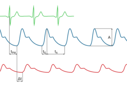
Additional key parameters and indicators can be derived from recorded pulse waves and ECG tracks: Amplitude A: The height from the beginning of the steepest rise to the highest point of the pulse curve is the amplitude. Oscillometric Index: The oscillometric index marks the pressure stage where the highest amplitude was measured and is comparable to […]

Read More
PWI™ – Pulse Wave Index

PWI™ – Pulse Wave Index

The PWI™ (Pulse Wave Index) is an indicator that relies on characteristics of the pulse wave shape (pulse amplitude and rise time) rather than on pressure values and its calculation is independent of the blood pressure. Studies suggest that, if taken together with the oscillometric ABI, it is the indicator with the highest sensitivity to determine […]

Read More
TBI – Toe Brachial Index

TBI – Toe Brachial Index

The TBI (Toe-Brachial-Index) is a common and easy to measure indicator to objectively proof an arterial disease in the lower extremities. As recommended by most guidelines, it is applied complementary to an ABI test, especially if the results are very high (ABI > 1.4, indicating a media sclerosis) or unreliable. TBI tests can be especially […]

Read More
ABI – Ankle Brachial Index

ABI – Ankle Brachial Index

The ABI (Ankle-Brachial-Index) is an evaluation criterion of the perfusion in the extremities based on the pressures in the arteries. It is mainly used to determine blood flow disorders related to peripheral arterial occlusive disease (PAOD). Historically, the systolic blood pressures are taken manually on each extremity individually by applying a pressure cuff and a […]

Read More